

Article Figures & Data
Figures
- Figure 1.
Development of vasculature scaffold in the RMS. A, Micrographs displaying BVs in the RMS at different developmental periods. Blood vessels were labeled by PECAM immunostaining, and RMS was visualized by dense DAPI labeling. Note that the first parallel BVs (arrows) appear at the outer border of the RMS. Note also that RMS width is decreasing from P3 to P60. RMS is indicated by the dashed white line. B, Quantification of the total RMS volume, from the OB until SVZ, at different developmental periods. C–E, Quantification of BVs density (C), the density of parallel BVs (D) and the number of BV branching points normalized to the density of BVs (E) at different developmental periods. At early developmental periods, the parallel BVs are longer at the outer border of the RMS. F, Proliferating endothelial cells in the RMS at the beginning of postnatal development. Proliferating endothelial PECAM+ cells were revealed by BrdU immunostaining, with BrdU being injected 1 h before animal perfusion. Orthogonal projection of a BrdU+/PECAM+ cell is presented as viewed in the x–z (bottom) and y–z (right) planes. Note that proliferating endothelial cells (colocalization of PECAM/BrdU) were mostly found at the borders of the RMS. In contrast to early periods of development (P3), no angiogenesis was detected in adulthood (P60). G, Migrating endothelial “tip” cells with filopodia in the developing RMS visualized by PECAM immunostaining. H, Micrographs showing nonfunctional BVs (arrows) at early stages of RMS development (P3). Colocalization between PECAM and dextran Texas Red indicates functional blood-carrying vessels. Note the presence of PECAM-positive but dextran Texas Red-negative nonfunctional blood vessels (arrows) in P3 RMS and their complete absence in the adult migratory stream. RMS is indicated by the dashed white line. I, Quantification of nonfunctional blood vessels in the RMS at different developmental periods. The values are presented as a percentage of length of PECAM+/dextran Texas Red− blood vessels from the total length of blood vessels.
- Figure 2.
RMS astrocytes regulate the formation and growth of BVs A, Micrographs depicting PECAM+ BVs (red) and GFAP+ astrocytes (green) at the beginning of postnatal development (P3, top) and in adulthood (P60, bottom). Note that at the early developmental periods, astrocytes are positioned at the outer border of the RMS, whereas in adults they are located inside of the migratory stream. RMS is indicated by the dashed white line. B, In vitro method for studying the role of astrocytes on BV formation and growth in cultures. Astrocytes were prepared from the different brain regions of P3 pups or from adult SVZ, and the ring of aorta was taken from adult mice. The conditioning medium (CM) derived from astrocytes was added to a 3D cultured aorta's rings every second day. C, Confocal image of cultured astrocytes. Astrocytes were stained with GFAP antibodies (green), and the cell nuclei were depicted by DAPI staining (red). Note that almost every DAPI-positive cell is colocalizing with GFAP, indicating high purity of astrocyte cultures. D, Quantification of cell density and purity of astrocyte cultures prepared from cortex, cerebellum, and SVZ-RMS. The first graph shows that there is no difference in the cell number of cultured astrocytes derived from different brain regions. The second graph shows the high purity of astrocyte cultures. E, Representative images showing BV growth from the aortic ring in the absence (control) and presence of conditioning medium derived from astrocytes of different regions. Arrows indicate newly formed BVs. F, Formation and growth of new blood vessels from the aortic ring after addition of conditioning medium from cortical, cerebellar, and SVZ astrocyte cultures. Note that only conditioning medium from P3 SVZ astrocytes potentiates formation and growth of new blood vessels. G, Western blot analysis of astrocyte culture-derived conditioning medium (CM) showing that in contrast to P3 cortex, P3 cerebellum, and adult SVZ-RMS, P3 SVZ-RMS-derived astrocytes secrete VEGF. Since adult astrocytes require longer culturing to reach the confluence, we have also immunoblotted conditioning medium of 14 d cultured adult SVZ astrocytes. Astrocyte cultures from which conditioning medium was derived were also immunoblotted for GFAP to ascertain an equal amount of astrocytes used for experiment.
- Figure 3.
RMS astrocytes express VEGF in vivo and in vitro. A, VEGF mRNA expression pattern in P3 RMS. Note the high density of VEGF-expressing cells at the RMS borders (arrows) and absence of signal in the center of the migratory stream. B, C, Fluorescent in situ hybridization for VEGF (red) in the cortex (B) and RMS (C) of P3 GFAP-GFP mice (astrocytes in green). Note the VEGF mRNA expression by cortical neurons and not by astrocytes in P3 cortex. In contrast, in P3 RMS astrocytes express a high level of this trophic factor. D–G, Micrographs showing a VEGF protein expression pattern in vivo in the RMS (D, top, E) and cerebellum (G) in P3 GFAP-GFP mice and in vitro in SVZ-RMS astrocytes cultures (F). In the RMS, VEGF is expressed by astrocytes (colocalization of VEGF and GFP). D, Note that VEGF is expressed mostly at the borders of P3 RMS (top) where the astrocytes are located and where the first parallel BVs appear. VEGF expression is drastically reduced at P14 when astrocytes appear in the center of the RMS (bottom). E, Micrographs are higher-magnification images showing expression of VEGF by P3 RMS astrocytes. A different optical section of astrocyte-expressing VEGF is also shown (bottom). G, In contrast to RMS, in the cerebellum VEGF is mostly expressed by neurons. H, Western blot analysis for VEGF, GFAP, and actin in P3, P7, P14, and adult RMS samples. Note that VEGF expression is markedly decreased from P3 to P7 until adulthood. I, Quantification of VEGF expression level during RMS development. The data were normalized to the level of GFAP, and P3 values were taken as 100%.
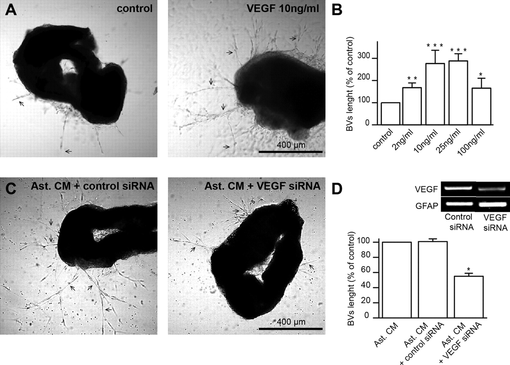
- Figure 4.
VEGF gain- and loss-of-function experiments affect the vasculature development. A, Representative images showing the newly formed BVs (arrows) in 3D cultures of aortic rings under control conditions and after application of exogenous VEGF (10 ng/ml). B, Quantification of the length of newly formed BVs under the control condition and after application of VEGF at different concentrations. C, Representative images of aortic rings cultured with the conditioning medium derived from P3 SVZ astrocyte culture (Ast. CM) transfected with control and VEGF siRNAs. D, Quantification of BV length reveals almost 50% reduction in cultures treated with conditioning medium derived from VEGF siRNA-transfected astrocytes compared with control conditions with conditioning medium derived from untransfected or control siRNA-transfected astrocytes. Inset, PCR analysis for VEGF and GFAP from astrocyte cultures transfected with control or VEGF siRNAs.
- Figure 5.
RMS-SVZ astrocytes can be specifically targeted in vivo. A, Confocal images demonstrating the high infection level after GFAP-GFP VEGF miRNA injection into two sites of P3 RMS. Infected cells are in green, and RMS was delimited by DAPI labeling (blue). B, High-magnification images demonstrate absence of colocalization between Dcx (red) and GFP-infected (green) cells. C, High-magnification images showing the colocalization of GFP-infected cells (green) with GFAP+ astrocytes (red). D, Micrograph showing colocalization between GFP (green) and GFAP (red). Orthogonal projection of a GFP+/GFAP+ cell is presented as viewed in the x–z (bottom) and y–z (right) planes. E, F, Injection of lentivirus expressing ZsGreen protein under the GFAP promoter into GFAP-GFP or GAD67-GFP RMS to ascertain the specific infection of astrocytes with the virus having GFAP promoter. The immunostaining with antibodies against GFP (red) and ZsGreen (blue) show that GFAP-ZsGreen virus infects only GFAP+ astrocytes (F) and not GAD67+ neuroblasts (E).
- Figure 6.
VEGF downregulation in RMS astrocytes alters the vasculature development in vivo. A, Profile of RMS vasculature at P15 after stereotaxic injection of control and VEGF miRNA viruses with GFAP promoter at P4. BVs were filled by dextran Texas Red (red). The viruses also drive GFP expression, allowing easy identification of infected cells (green). Nuclear DAPI staining (blue) is delimiting the RMS, which can be distinguished by the high density of cells. Note that with GFAP-GFP control miRNA, infected RMS contains more long and parallel BVs (arrows) with fewer sprouts compared with RMS infected with VEGF miRNA. Inset, Western blot analysis of VEGF downregulation in VEGF miRNA-injected RMS compared with the control miRNA-injected mice. Only the RMS regions containing GFP+-infected cells were dissected and used for immunoblotting. The data were normalized to GFAP level. B–D, Quantification of overall BV density (B), the density of parallel BVs (C), and BV branching points (D) in the RMS and at its border at P15 after infection with control and VEGF miRNA viruses with GFAP promoter.
- Figure 7.
Neuroblast migration is less efficient at the beginning of postnatal development. A, Representative confocal images of retrovirally labeled neuroblasts (green) and BVs filled with dextran Texas Red (red) in P5, P14, and adult RMS showing the distribution of neuroblasts at the RMS center and border and their distance from BVs. Note that at P5 and P14, neuroblasts located at the RMS borders are closer to the BVs compared with those in the core of RMS. B, Summary graphs showing the distribution of the shortest distance of neuroblasts from BVs at different developmental stages. Note that at P5 and P14, over 80% of neuroblasts in the RMS border are located <5 μm away from BVs. In contrast, in the RMS center, the distance between neuroblasts and BVs is much bigger and the distribution of the shortest distance of neuroblasts to BVs is more homogeneous. C, Real-time videoimaging of retrovirally labeled neuroblast (green) migrating on dextran Texas Red filled BV (red) in the acute slices of P5 mouse forebrain. D, Time-lapse videoimaging of migrating neuroblasts during RMS postnatal development. Slices were derived from P4/P5, P13–P15, and adult mouse forebrain. Migrating cells are indicated during their migratory (arrows) and stationary (arrowheads) phases. E, Migration distance of neuroblasts at different developmental stages. Note that at the early developmental periods, the displacement of neuronal precursors is significantly smaller. F, Total distance propagated by neuronal precursors in the RMS at different developmental stages. Note that migration at the border of P5 and P14 RMS is more efficient than in the center of RMS. G, Rate of neuroblast migration in the RMS at different developmental stages. Although the rate of neuroblasts migration in P5 RMS is significantly smaller compared with P14 and adult RMS, no differences in the RMS subregions were observed. H, Summary graph showing differences in the duration of stationary periods across the RMS subregions and developmental stages.
- Figure 8.
VEGF downregulation at the early postnatal developmental stages affects the migration of neuroblasts. A, Time-lapse videoimaging of mCherry+ migrating neuroblasts (red) at P15 after stereotaxic injection of control or VEGF miRNA lentiviruses with GFAP promoter (green) at P4 RMS. Migrating cells were labeled by stereotaxic injection of mCherry-expressing lentiviruses into P10 SVZ. Migrating cells are indicated during their migratory (arrows) and stationary (arrowheads) phases. B, Summary graph illustrating that the migration distance of neuroblasts is significantly decreased when the astrocytes were infected with VEGF miRNA. C, Total cell displacement of neuroblasts in P15 RMS after stereotaxic injection of control or miVEGF lentiviruses at P4. Note the reduction in the cell displacement after downregulation of VEGF in RMS astrocytes. In contrast, no changes in the migration of neuronal precursors were observed after acute VEGF application, suggesting that this trophic factor does not have any direct role on neuroblasts migration. D, Unaltered rate of neuroblast migration in the RMS after VEGF downregulation in astrocytes at early developmental periods and after acute VEGF application. E, Summary graph illustrating that the duration of the stationary periods is significantly higher in the RMS where VEGF was downregulated in the astrocytes. In contrast, acute VEGF application does not induce any effect on neuroblasts migration. F, Representative images of RMS explants (top) cocultured with astrocytes infected either with control or VEGF miRNA. The miRNAs viruses also express GFP, which allows the easy identification of infected cells (bottom). No difference in the length of neuroblast chains arising from explants cocultured with control or VEGF miRNA was observed. G, Graphs showing the distribution of shortest distance of neuroblasts from BVs in control, VEGF miRNAs-injected animals, and in naive (noninjected) mice. miRNAs were injected at P3, and mCherry-expressing lentivirus was injected into the SVZ at P12. Note that the distance between neuroblasts and BVs is increasing when VEGF is downregulated in the RMS astrocytes. H, Micrographs illustrating the reduced number of BrdU+ cells in the GCL of the OB after alteration in the vasculature scaffold by VEGF downregulation in the RMS astrocytes. I, Summary graphs illustrating a decrease in the density of BrdU+ cells in GCL of OB and an accumulation of neuronal precursors in the RMS in VEGF miRNA-injected mice. Altered distribution of BrdU+ cells along SVZ–OB pathway suggests affected migration of neuronal precursors.


