

Article Figures & Data
Figures
- Figure 1.
Generation of GEFS+ and control flies by targeted knock-in. A, Schematic diagram of a sodium channel α-subunit. Lysine residue altered in the human K1270T GEFS+ mutation is located in the second transmembrane segment in homology domain III (indicated by an asterisk). Comparison of the amino acid sequence in this region reveals a high degree of homology between species. Identical amino acids are shaded gray. Location of lysine residue altered in mutants is indicated by the asterisk. B, Diagram of the wild-type para sodium channel locus and the targeting vectors. The GEFS+ targeting vector contained the K to T substitution, and the control targeting vector contained the control substitution K to K. Location of the substitution is indicated by an asterisk. Homologous recombination events between incoming linear recombinogenic DNA and endogenous para locus are indicated by crossed lines.
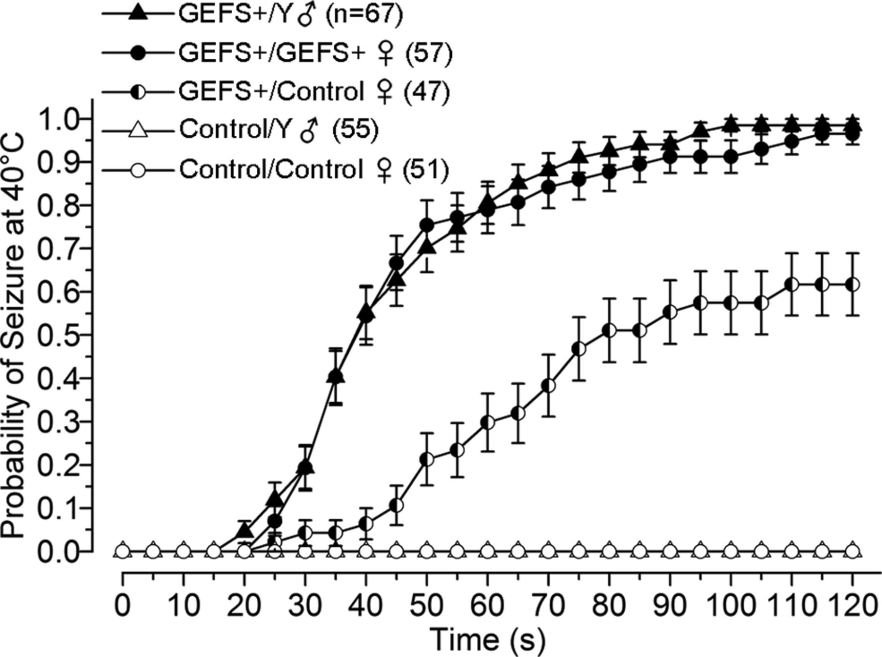
- Figure 2.
GEFS+ flies exhibit a temperature-induced seizure phenotype. Seizures are defined as a brief period of leg twitches, followed by an inability to maintain standing posture, with wing flapping, leg twitching, and sometimes abdomen curling. Individual 2-d-old flies were put into vials that were immersed in a water bath (40°C, 2 min). The status of each fly (seizing or not seizing) was determined at 5 s intervals, and the probability of seizing at each time point was calculated for the population of flies examined in each genotype. The probability of seizing after 2 min at 40°C is significantly different between the mutants (GEFS+/Y and GEFS+/GEFS+), heterozygotes (GEFS+/control) and controls (control/Y and control/control; p < 0.001, ANOVA, Bonferroni's post hoc test). Symbols and error bars represent mean ± SEM from the number of flies indicated (n).
- Figure 3.
Sodium currents in GEFS+ and control LNs. A, A LN in the dorsal lateral cluster in the antennal lobe, filled with biocytin during recording, in a control and GEFS+ brain. Confocal images of the brains fixed and double stained with a fluorescently labeled secondary antibody to biocytin and Nc82 (anti-bruchtpilot antibody) are shown. Arrows indicate the LN cell body (control) or axon initial segment (GEFS+). The soma of the GEFS+ cell was lost during pipette removal. B, Depolarizing voltage-step-elicited sodium currents at room temperature (23°C) and following heating of the recording solution to high temperature (35°C). INaP in control LNs decayed to baseline after the pipette potential returned to −75 mV, but it remained activated in the GEFS+ LN at 35°C (arrow). Sodium currents could not be clamped in either genotype. C, INaT amplitudes are no different in GEFS+ and control LNs at 23 or 35°C (independent t test, 23°C, p = 0.28; 35°C, p = 0.07), and the amplitude did not change with temperature in either genotypes (paired t test, control, p = 0.08; GEFS+, p = 0.20). D, The voltage step required to elicit the first inward sodium current is significantly more hyperpolarized in GEFS+ than in control at both 23 and 35°C. ***p < 0.001 (independent t test). The current activation threshold did not change significantly with temperature in either genotype (paired t test, control, p = 0.06; GEFS+, p = 0.14). Symbols and error bars represent mean ± SEM.
- Figure 4.
Temperature-induced hyperpolarizing shift in INaP deactivation threshold is significantly larger in GEFS+ LNs. A, B, Families of INaP associated with the variable test step during the three-step voltage protocol illustrated in a typical control and GEFS+ LN at 23 and 35°C. Only one trace during the prepulse is shown for clarity. The first test potential that results in decay of the current back to baseline is defined as the current deactivation threshold voltage and is indicated by the arrow in each set of currents. When the temperature is raised from 23 to 35°C, current deactivation voltage shifts from −45 to −55 mV in control and −55 to −75 mV in GEFS+ LNs. Ai, Bi, I--V curves representing INaP amplitude measured during last 10 ms of the test pulse for each test potential from the traces shown above. Dashed lines are linear fits used to calculate the conductance of the INaP. C, The deactivation voltage is more hyperpolarized at 35°C compared to 23°C in both control and GEFS+. However, the deactivation voltage is significantly more hyperpolarized in GEFS+ than in control at 35°C. D, Mean INaP conductance reversibly increase at 35°C in both genotypes but was not different between GEFS+ and control at 23°C (independent t test, p = 0.29) or 35°C (p = 0.38). *p < 0.05 (independent t test); **p < 0.01, ***p < 0.001 (paired t test). E, Average activation and deactivation threshold voltage of sodium currents in control and GEFS+ at room temperature and elevated temperature. GEFS+ LNs have a wider voltage range over which sodium channels are conductive. Symbols and error bars represent mean ± SEM.
- Figure 5.
Alterations in evoked firing properties of GEFS+ LNs at elevated temperature. A, B, Representative trains of spikelets recorded from control and GEFS+ LNs at 23 and 35°C evoked by the stimulus protocol illustrated. Ai, Bi, Spikelet frequency plotted as a function of injected current for LNs shown above. C, The incidence of poststimulus depolarization is defined as the percentage of traces in which there was a poststimulus depolarization that lasted for at least 50 ms in each LN. The incidence of GEFS+ LNs showing poststimulus depolarizations increased significantly at 35°C compared to 23°C (paired t test, *p < 0.05). In contrast, the incidence of poststimulus depolarization was low in control LNs and did not increase significantly at elevated temperature (p = 0.36). D, Raising the temperature from 23 to 35°C resulted in a significant increase in the maximal firing frequency in control LNs and a significant decrease in GEFS+ LNs. *p < 0.05 (paired t test). Symbols and error bars represent mean ± SEM.
- Figure 6.
Appearance of sustained depolarizations without spikelets in GEFS+ LNs at high temperature. A, B, Continuous recordings of spontaneous activity in control and GEFS+ LNs as the temperature is raised and lowered as indicated by the overlay of the temperature probe recording. Ai–Aiii, Bi–Biii, Traces on expanded time scales from the regions indicated. In both genotypes, the regular burst firing frequency gradually decreases as the temperature increases. Before cessation of firing, activity in GEFS+ LNs at high temperature is characterized by the appearance of sustained depolarizations without spikelets that are not observed in control LNs. Normal burst firing resumes in both genotypes when the bath is returned to room temperature. C, The frequency of sustained membrane depolarizations in GEFS+ LNs is increased at high temperature. These events were counted at 23°C (5 min before heating), at high temperature (30–35°C), and following return to room temperature (0–5 min after return to room temperature). D, The percentage of membrane depolarizations that were sustained increased at 35°C in GEFS+. E, Spikelet frequency increased in control LNs, but decreased in GEFS+ LNs at 35°C compared to 23°C. F, The spikelet threshold is more hyperpolarized in GEFS+ than in control LNs at both 23 and 35°C (independent t test, p < 0.05). The spikelet threshold did not change as a function of temperature in either GEFS+ (paired t test, p = 0.95) or control LNs (p = 0.23). *p < 0.05 (paired t test). Symbols and error bars represent mean ± SEM.
- Figure 7.
PTX increases GEFS+ sensitivity to heat-induced seizures. A, Flies were fed sucrose with indicated concentration of PTX and tested for seizure susceptibility within 30 min. The probability of seizing in GEFS+ flies after 2 min at 37°C increases with PTX concentration (p < 0.001, one-way ANOVA). Control flies fed the maximal dose of PTX (0.4 mm) did not show seizure behavior at 37°C. B, In GEFS+ flies, the time spent seizing during the 2 min heat exposure increases with PTX concentration (p < 0.001, one-way ANOVA). Symbols and error bars represent mean ± SEM.


