

Article Figures & Data
Figures
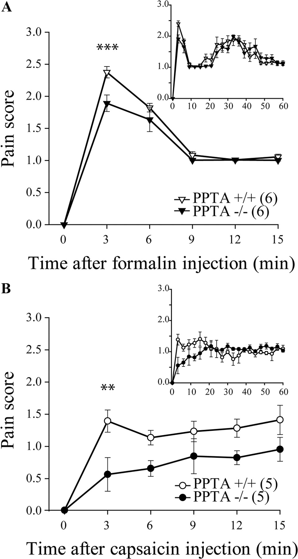
- Figure 1.
Formalin- and capsaicin-induced pain behaviors in PPTA−/− mice. PPTA−/− and their PPTA+/+ littermates were injected with formalin or capsaicin in the plantar surface of the hindpaw, and their pain behaviors were observed for 60 min (A, B, insets). A, Intraplantar formalin (10 μl of 5.4% formalin) produced a significant increase in pain-related behaviors in PPTA+/+ mice, whereas the maximal pain score was lowered in PPTA−/− mice during the first few minutes after injection. ***p < 0.001, two-way ANOVA with Bonferroni's post hoc test. B, Subcutaneous capsaicin (6 μg in 10 μl) produced a significant increase in pain-related behaviors in PPTA+/+ mice, an effect that was reduced in PPTA−/− mice over the first 15 min of the test. **p < 0.01, two-way ANOVA with Bonferroni's post hoc test. The numbers in parentheses represent the number of animals per group. Error bars indicate SEM.
- Figure 2.
Inhibition of formalin-induced pain behavior following activation of DOPR and MOPR. Sprague Dawley male rats were injected with intradermal formalin (50 μl of 5.4%) in the plantar surface of the hindpaw, and pain behaviors were recorded during 60 min. A, Unlike intraplantar injection of saline, intraplantar injection of formalin produced a biphasic nociceptive response. Intrathecally administered deltorphin II (10 μg; 12.7 nmol) produced a decrease in formalin-induced pain behaviors. NTI (50 nmol) reversed the effects of Dlt II. B, Intrathecally administered DAMGO (1 μg; 1.9 nmol) produced a decrease in formalin-induced pain behaviors compared with saline (for reference purpose, results presented are the same as in A). CTOP coinjection (6 nmol) reversed the effects of DAMGO. C, Graphic representation of AUC for the first 15 min (highlighted by a gray area in A and B) was calculated from data presented in A and B. *p < 0.05, **p < 0.01, and ***p < 0.001, one-way ANOVA with Bonferroni's post hoc test. The numbers in parentheses represent the number of animals per group. Error bars indicate SEM.
- Figure 3.
Reduction of formalin-induced neuronal activation by DOPR and MOPR agonists. Sprague Dawley male rats were injected with intradermal formalin (50 μl of 5.4%) in the plantar surface of the hindpaw, and c-fos expression in the spinal cord was observed by immunohistochemistry. When intradermal vehicle was injected, little c-fos expression was observed (A), whereas formalin induced robust c-fos expression (B). C, Rats treated with intrathecal Dlt II (10 μg; 12.7 nmol) 5 min before formalin displayed a significant reduction in c-fos expression. D, NTI (50 nmol) reversed the effects of Dlt II. E, Intrathecal DAMGO (1 μg; 1.9 nmol) also produced a reduction in c-fos expression, and this effect was suppressed by CTOP (6 nmol) (F). CTOP also induced an increase in c-fos expression in deeper laminae (F). G, Graphic representation of the number of c-fos-positive neurons. *p < 0.05 and ***p < 0.001, two-way ANOVA with Bonferroni's post hoc test. The numbers in parentheses represent the number of animals per group. Error bars indicate SEM.
- Figure 4.
Inhibition of capsaicin-induced pain behaviors following activation of DOPR and MOPR. Sprague Dawley male rats were injected with subcutaneous capsaicin (15 μg in 50 μl) in the plantar surface of the hindpaw, and pain behaviors were recorded from 0 to 60 min. The first 15 min were analyzed further. A, No pain behavior was noted in rats treated subcutaneously with vehicle. Conversely, subcutaneous injection of capsaicin produced pain behaviors that were decreased by intrathecally administered deltorphin II (10 μg; 12.7 nmol). NTI preinjection (50 nmol) reversed the effects of Dlt II. B, Intrathecally administered DAMGO (1 μg; 1.9 nmol) produced a decrease in capsaicin-induced pain behaviors. CTOP preinjection (6 nmol) reversed the effects of DAMGO, compared with saline (results presented are the same as in A). C, Graphic representation of AUC obtained from the first 15 min of A and B. *p < 0.05 and ***p < 0.001 with Bonferroni's post hoc test. The numbers in parentheses represent the number of animals per group. Error bars indicate SEM.
- Figure 5.
Reduction of capsaicin-induced neuronal activation by DOPR and MOPR agonists. Sprague Dawley male rats were injected with capsaicin (15 μg in 50 μl) in the plantar surface of the hindpaw, and c-fos expression in the spinal cord was observed by immunohistochemistry. When subcutaneous vehicle was injected in the paw, little c-fos expression was observed (A), whereas capsaicin induced robust c-fos expression (B). C, When rats were treated intrathecally with Dlt II (10 μg; 12.7 nmol) 5 min before capsaicin injection, a significant reduction in c-fos expression was observed. D, NTI preinjection (50 nmol, 5 min before Dlt II) reversed the effects of Dlt II but also increased c-fos expression in deeper laminae. E, Intrathecal DAMGO (1 μg; 1.9 nmol) also produced a reduction in c-fos expression, and its effect was suppressed by a preinjection of CTOP (6 nmol) (F). G, Graphic representation of the number of c-fos-positive neurons. *p < 0.05, **p < 0.01, and ***p < 0.001, two-way ANOVA with Bonferroni's post hoc test. The numbers in parentheses represent the number of animals per group. Error bars indicate SEM.
- Figure 6.
Formalin- and capsaicin-induced NK1R internalization. NK1R internalization in the spinal cord of male Sprague Dawley rats was observed by immunofluorescence. A, In control rats, NK1R-like immunostaining was clearly visible throughout the superficial laminae of the spinal cord. Immunolabeling of NK1R appeared at the cell surface. Ten minutes following intradermal injection of formalin, NK1R immunostaining in the ipsilateral side appeared as being inside the cells in vesicle-like structures (C). By contrast, NK1R labeling in the contralateral side of the spinal cord remained at the cell surface (B). When Dlt II (30 μg; 38.1 nmol) was injected intrathecally 5 min before formalin, a significant reduction in NK1R internalization was observed (D). This effect was blocked by NTI (150 nmol) (E). A similar effect was seen when DAMGO (10 μg; 19.4 nmol) was injected intrathecally 5 min before formalin (F), an effect blocked by CTOP (30 nmol) (G). Similarly, capsaicin induced a robust NK1R internalization in the ipsilateral side of the spinal cord (I) but not on the contralateral side (H). Injection of Dlt II (J) or DAMGO (L) 5 min before capsaicin injection prevented NK1R internalization. Again, the effects of Dlt and DAMGO were blocked by preinjection of NTI and CTOP, respectively (K, M). N, Graphic representation of the percentage of neurons with internalized NK1R induced by formalin and capsaicin injections for ipsilateral and contralateral sides of the lumbar spinal cord. *p < 0.05, **p < 0.01, and ***p < 0.001, one-way ANOVA with Bonferroni's post hoc test. The numbers in parentheses represent the number of animals per group. Scale bar: B, 30 μm. Error bars indicate SEM.
- Figure 7.
SP-induced NK1R internalization. The effects of Dlt II or DAMGO on exogenous SP-induced NK1R internalization were verified by immunofluorescence. In control rats, no NK1R internalization was observed in a majority of neurons (A), whereas intrathecal injection of SP (22 nmol) induced robust NK1R localization in intracellular, vesicle-like structures (B). Neither intrathecal Dlt II (30 μg; 38.1 nmol) nor DAMGO (10 μg; 19.4 nmol) treatment 5 min before SP injection was able to block the SP-induced NK1R internalization (C, D, respectively). Scale bar: A, 20 μm.


