

Article Figures & Data
Figures
- Figure 1.
Transgenic lines that mark RGC subsets. a , Transgenic lines used in this study. b , c , e , f , Portions of retinas showing YFP-marked J-RGCs in a JxTSY mouse ( b ), BD-RGCs in a BDxTSY mouse ( c ), W3-RGCs in a TYW3 mouse ( e ), and W7-RGCs in a TYW7 mouse ( f ). d , g , Retinas from a BDxTSY mouse ( d ) and a TYW7 mouse ( g ) with reduced density of labeling resulting from manipulation of Cre as shown in a and described in results. Scale bar: (in g ) b–g , 100 μm.
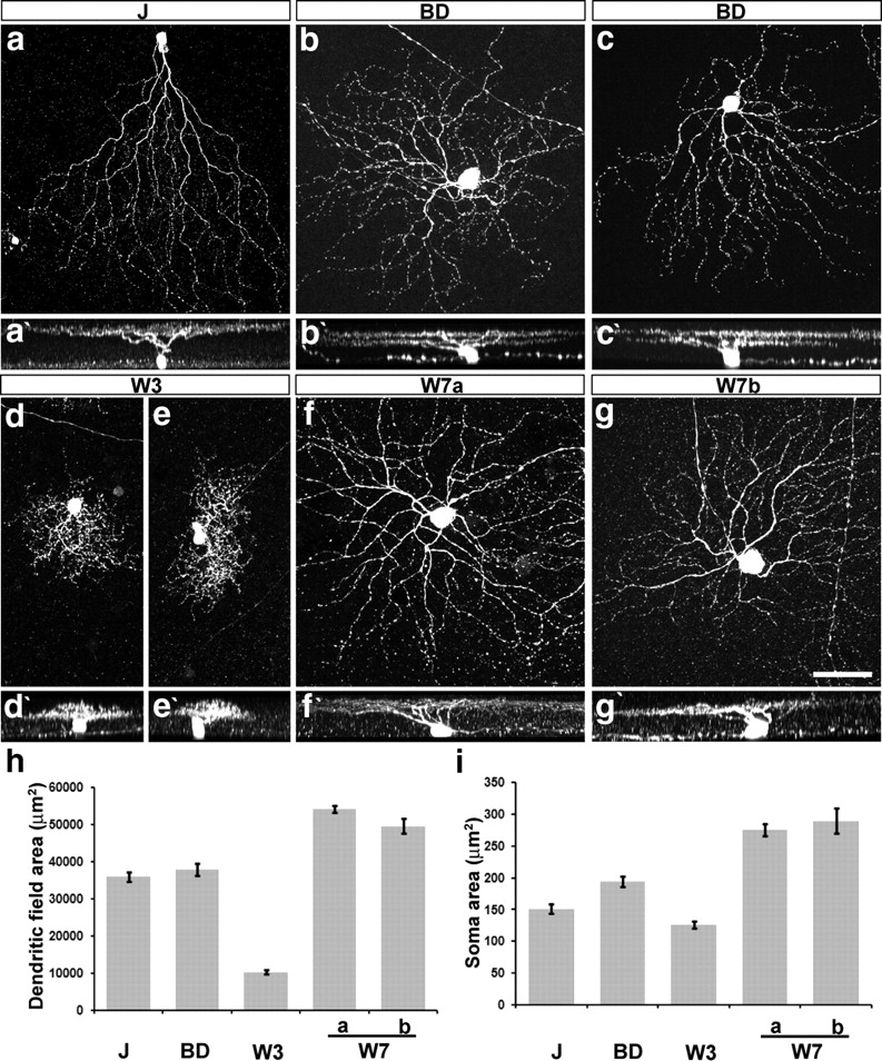
- Figure 2.
Morphology of J-, BD-, W3-, and W7-RGCs. a–g , Confocal stacks of J-, BD-, W3-, and W7-RGCs. W7a and W7b indicate RGCs with dendrites in SL1–SL2 and SL4 or only in SL4, respectively. a′–g′ , Z rotations of cells shown in a–g . h , i , Dendritic field and soma areas of labeled RGCs, measured from images such as those in a–g (n = 13–27 cells per type). Error bar indicates SEM. Scale bar: (in g ) a–g , 50 μm.
- Figure 5.
Subtype-specific patterns of dendritic development in the inner plexiform layer. Retinal sections were from P5, P8, and P12–P13 mice. Staining and abbreviation are as in Figure 3. All subtypes have lamina-restricted dendrites by P12–P13 ( c , f , i , l ; compare with Fig. 3), but the extent and nature of postnatal remodeling varies among subtypes. Scale bar: (in l ) a–l , 20 μm.
- Figure 6.
Lamina-restricted arbors of RGC axons within the retinorecipient zone of the superior colliculus. a–j , Vibratome sections were immunostained with anti-GFP (green) and counterstained with Neurotrace or TO-PRO (blue). a , c , e , g , i , Coronal sections from mice that had been monocularly enucleated to ablate retina axons from the side marked “ipsi.” b , d , f , h , j , Sagittal sections from other mice that had not been enucleated. Brackets indicate the span of axonal arbors of RGC subtypes. Transgenic lines are indicated at the left; mice were 40 d of age or older. k , The entire retinorecipient zone is marked by injection of cholera toxin b subunit (CTB, red) into one eye, and corresponds to that marked in Chx10-Cre × TSY mice in a . Arrows in a and k show sparse ipsilateral projections, which terminate in a deep region. l , Schematic showing distinct axonal restriction of each type of RGCs and ipsi projection. Scale bar: (in k ) a–k , 300 μm.
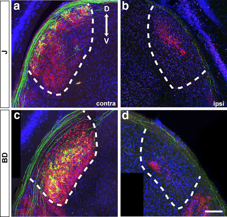
- Figure 7.
Lamina-restricted arbors of RGC axons in the lateral geniculate nucleus. Coronal sections were stained with anti-GFP (green) and Neurotrace (blue) after enucleation and cholera toxin administration (CTB, red). a , b , J-RGCs. c , d , BD-RGCs. Cholera toxin-positive regions in b and d contain projections from the ipsilateral retina. D, Dorsal, V: ventral. Scale bar: (in d ) a–d , 150 μm.
- Figure 8.
Subtype-specific patterns of axonal development in the superior colliculus. Sagittal sections from P5–P6, P8, P11–P13, and P17–P18 mice are shown. Axonal arborization from each RGC subtype was visualized by immunostaining to anti-GFP (green). i′–p′ , High magnification of boxes marked in i–p to emphasize distinct patterns of axonal arborization in W3- and W7-RGCs. The pia surface is indicated by a red line. Blue, Cell bodies. Scale bars: (in p ) a–p , 200 μm; (in p′ ) i′–p′ , 100 μm.
- Figure 9.
Subtype-specific patterns of axonal development in the lateral geniculate nucleus. Sections were immunostained by anti-GFP (green) at P5–P6, P7–P8, and P11. The dotted lines mark the boundary of LGN. Scale bar: (in f ) a–f , 100 μm.
- Figure 10.
Summary of subtype-specific patterns of dendritic and axonal development. See Results for details.


Tables
- Table 1.
Correspondence between RGC subsets identified by transgenic markers and those categorized by morphological criteria
This study Sun et al. (2002) Coombs et al. (2006) Kong et al. (2005) Badea and Nathans (2004) Völgyi et al. (2009) J C6 M5a — 6 G15 BD D1, D2 12, 13 — Bistratified 1, 2 G16, G17 W3 B2 M11 1 4 G5 W7 A2 M9 10 7 G3


