

Article Figures & Data
Figures
- Fig. 1.
The human D1 dopamine receptor promoter–cholera toxin (D1CT) fusion transgene. E, EcoRI;N, NotI; (N),NotI site deleted during cloning; ORF, open reading frame (coding sequence); S,SalI; SD/SA, splice donor/splice acceptor sites; UT, untranslated region.
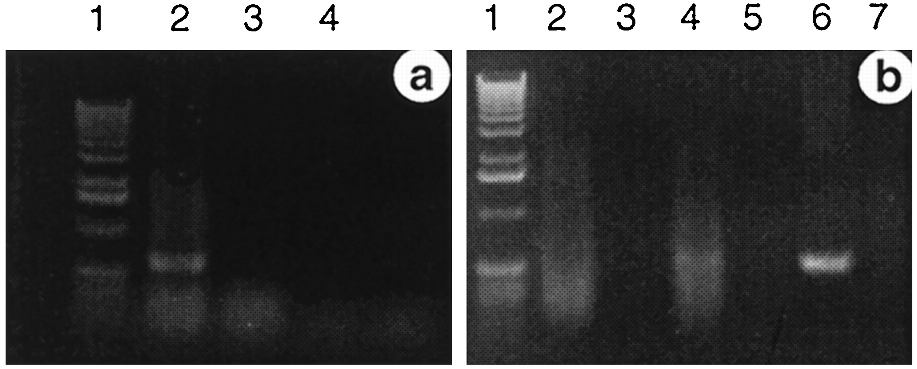
- Fig. 2.
Detection of cholera toxin mRNA in brain extracts of affected D1CT mice. a, RT-PCR of affected D1CT-11 mouse brain poly(A+) RNA. Lane 1, Kilobase ladder DNA size marker; lane 2, RT included (+RT); lane 3, RT omitted (−RT); lane 4, RNA omitted (−RNA). b, RT-PCR of nontransgenic (control) littermate and affected D1CT-7 mouse brain poly(A+) RNA. Lane 1, Kilobase ladder DNA size marker; lane 2, control, +RT; lane 3, control, −RT; lane 4, D1CT-7, +RT;lane 5, D1CT-7, −RT; lane 6, D1CT-7 genomic DNA template (positive control); lane 7, −RNA. CT mRNA is detected in the affected D1CT-11 and D1CT-7 mouse brain extracts as a 600 bp RT-PCR fragment (a, lane 2; b, lane 4) identical in size to a positive control CT genomic DNA PCR fragment (b, lane 6).
- Fig. 3.
D1CT transgene expression and D1 receptor and CNS neuroarchitecture of D1CT-7 mice. a, b, Bright-field view of ICC staining for the D1 receptor (dark staining with blue Nissl counterstain) in coronal brain sections, indicating no discernible changes in D1 receptor distribution, neuroanatomy, or density between control nontransgenic (a) and D1CT-7 (b) mice. c, d, Bright-field view of Nissl-stained sagittal brain sections, indicating no discernible changes in CNS morphology or neuron number between control nontransgenic (c) and D1CT-7 (d) mice. e, Bright-field view of ICC staining for the D1 receptor (dark staining) in a coronal section of the somatosensory and insular cortex [right (ventral to dorsal), insular, S2, S1] and the caudate-putamen (left), showing extensive D1 receptor expression in the caudate-putamen and less extensive but evident D1 receptor expression in the somatosensory and insular cortex, predominantly in layers II–III. f, Dark-field view of ISH staining for the presence of CT mRNA (light grains) performed on the same section shown in e, showing colocalization of CT mRNA with D1 receptor staining in the somatosensory and insular cortex layers II–III but not in the caudate-putamen. g, Dark-field view of ISH staining, detecting the presence of CT mRNA (light grains) in the piriform cortex layer II and the intercalated nucleus of the amygdala, regions that are also positive for D1 receptor mRNA and protein (data not shown). h, Higher magnification dark-field view of ISH staining of CT mRNA (light grains) in the intercalated nucleus of the amygdala in a coronal section parallel to that in g. Control nontransgenic littermates exhibit indistinguishable D1 receptor and Nissl staining in these regions and other CNS regions but no CT mRNA ISH staining (data not shown). This regionally restricted pattern of CT ISH staining to the areas shown was consistent between different D1CT-7 mice, reflecting the identical behavior of the animals of this line. For ISH, n = 3 per group; for Nissl, n = 4 per group; and for D1 receptor ICC,n = 2 per group. amy, Amygdala;c, cortex; CPu, caudate-putamen;en, endopiriform nucleus;ep/ic, entopeduncular nucleus/internal capsule; hpc, hippocampus; hyp, hypothalamus; icn, intercalated nucleus of the amygdala;pir, piriform cortex; I–VI, somatosensory cortical layers.
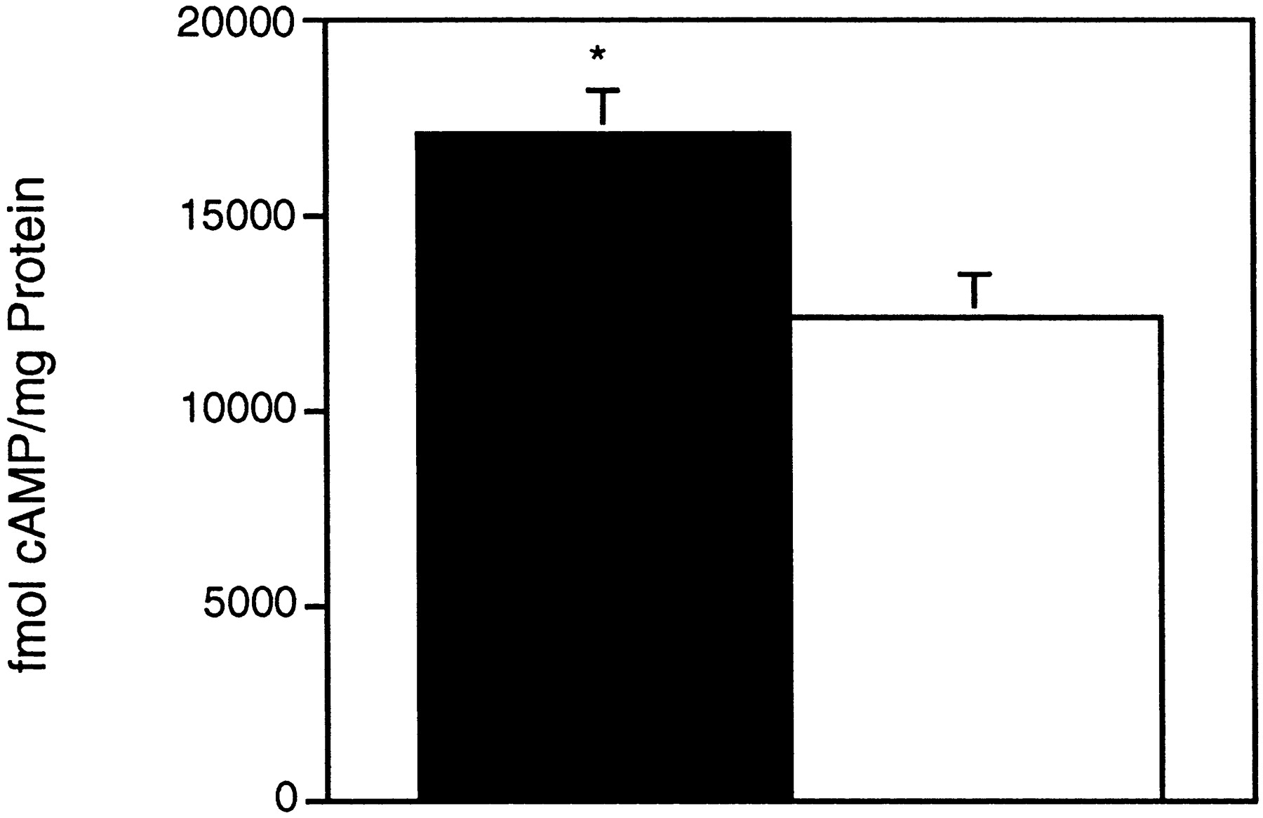
- Fig. 4.
Elevated cAMP levels in CT+ CNS regions of D1CT-7 mice. The mean cAMP content (femtomoles of cAMP per milligram of protein) of extracts prepared from whole dissected somatosensory and piriform cortex is shown. Filled bar, D1CT-7 mice;open bar, control nontransgenic littermate mice; error bars indicate +SEM; n = 6 mice per group; *p < 0.05 using Student’s ttest.
- Fig. 5.
D1CT-7 mice nonaggressively bite their sibling cage mates. a, Sibling missing tail.b, Sibling missing ears. c, The fraction of recorded audible distress vocalizations occurring while a D1CT-7 or control nontransgenic littermate mouse was observed to have its snout in contact with another mouse. Filled bar, D1CT-7 mice;open bar, control nontransgenic littermate mice; number of observation periods per mouse = 112; n = 3 mice per group; error bars indicate +SEM; ***p < 0.001 by Student’s t test. d, Resident–intruder aggression assay. Shown are the mean number of attacks by resident D1CT-7 transgenic mice or control nontransgenic littermates on an intruder mouse within 3 min. Filled bar, D1CT-7 mice; open bar, control nontransgenic littermate mice; n = 9 mice per group; error bars indicate +SEM; *p < 0.05 using repeated measures ANOVA [F(1,7) = 6.798;p = 0.035]. The outcome of the parametric analysis was confirmed by a Wilcoxon signed rank test (Z = −2.375; p = 0.018). e, Representative transgenic lineage of D1CT-7 mouse-biting behavior.Dark symbols, D1CT mice; open symbols, control nontransgenic littermate mice; gray symbol, mouse that died before determination of transgenic status;square, male; circle, female; /, bitten mouse; B, biting mouse (determined either by direct observation or by observation that it was the only unbitten mouse in the cage); (B), biting mouse (shown to bite mate when placed in a breeding cage). f, Sequential frames extracted from a video-recording of a D1CT-7 mouse (white) repeatedly biting a sibling mouse (agouti) during conspecific grooming rather than aggressive attack. Left, Initial grooming at face, 0 sec. Middle, Grooming behind head after several bites, 12 sec. Right, Bite and startle/vocalization of sibling, 12.5 sec. Biting while grooming continues in spite of vocalizations by the bitten mouse.
- Fig. 6.
Increased locomotion, gnawing, and leaping in D1CT-7 mice. A bar graph of the mean number of observed behaviors that were >3 consecutive seconds in duration (Fray et al., 1980) is shown.Filled bars, D1CT-7 mice; open bars, control nontransgenic littermate mice; n = 14 mice per group; error bars indicate +SEM; *p < 0.05, **p < 0.01, and ***p < 0.001 using Student’s t test. MANOVA of the predicted dopaminergically induced behaviors consisting of the dependent variables locomotion, rear, sniff, gnaw, groom, and leap indicated a significant difference between transgenic and control nontransgenic littermates [F(6,21) = 5.214;p = 0.002]. Therefore the aforementioned independent analysis of each variable was justified.
- Fig. 7.
Perseverative (long-duration) and repetitive behaviors in D1CT-7 mice. a–c, Representative behavioral waveform displays (Campbell et al., 1998) of normal behavior in individual control nontransgenic littermate mouse (a) and of perseverative behavior (b; in this instance, eating) or repetitive behavior (c; in this instance, locomote-rear) in D1CT-7 mice. The type of perseverative or repetitive behavior exhibited by the D1CT-7 mice continuously varies in each individual. In this display, behavioral activities analyzed are on the y-axis, whereas time is on the x-axis. Thus, vertical lines represent transitions from one behavior to another, whereas horizontal lines represent the duration of each behavior. Periods of rapid behavioral switching appear ascondensed vertical lines or bars.d, e, Comparison of the mean duration of all behaviors (d) or of nonlocomotor behaviors (e) exhibited by D1CT-7 and control mice.Filled bars, D1CT-7 mice; open bars, control nontransgenic littermate mice; n = 6 mice per group; error bars indicate +SEM; **p < 0.01 and ***p < 0.001 using Student’s ttest. d shows the mean duration of all behaviors, either simple stationary (1-state) or complex locomotor-dependent (2-state), observed within a 30 min period, whereas e shows the mean duration of all simple stationary (nonlocomotor-dependent) behaviors (i.e., eat, drink, self-groom, groom other, rear, dig, and bar hang) observed within a 30 min period.


